Fibroadenoma of the breast
Fibroadenoma is the most common benign breast tumor, mostly in young women. It consists in two components (epithelial and fibroblastic), estrogen-dependent, slowly growing.
Until recently, traditionally, fibroadenoma was considered to be a benign mixed tumor, but recent studies showed that only the fibroblastic component is neoplastic (being monoclonal), while the epithelial one is only reactive, non-neoplastic (being policlonal).

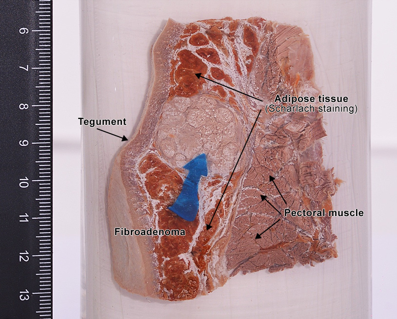
Figure 1
Gross examination : most often it is a solitary small sized (3 - 4 cm.) nodule, well circumscribed, firm, mobile (it doesnt infiltrate the teguments or the deep structures fat tissue or skeletal muscle) (Figure 1). On cut surface, the tumor is white-greyish, lobulated or cauliflower-like resemblance, with a whorl-like pattern and irregularly slit-like spaces.


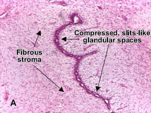
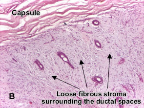
Microscopy : Fibroadenoma is nodular and encapsulated, included in breast. The epithelial proliferation appears in a single terminal ductal unit and describes duct-like spaces surrounded by a fibroblastic stroma. Depending on the proportion and the relationship between these two components, there are two main histological features : intracanalicular and pericanalicular. Often, both types are found in the same tumor.
Intracanalicular fibroadenoma (photo A) : stromal proliferation predominates and compresses the ducts, which are irregular, reduced to slits. Pericanalicular fibroadenoma (photo B) : fibrous stroma proliferates around the ductal spaces, so that they remain round or oval, on cross section. The basement membrane is intact. (H&E, ob. x4)
Additional information :

